A 33-year-old Chinese woman presented to the hospital with a 5-day history of fever and cough of unknown cause
She indicated that she worked in Wuhan, China (the center of novel coronavirus outbreak) but had traveled to Lanzhou, China, 6 days before presentation to the hospital.
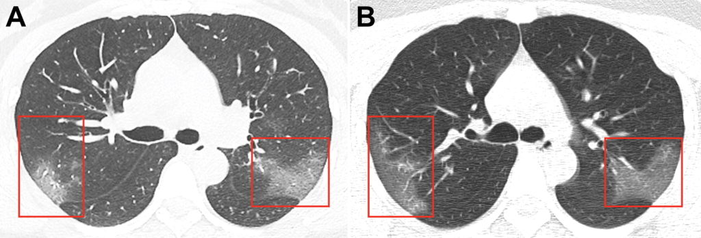
At admission, her body temperature was elevated to 39.0°C (102.2°F) and coarse breath sounds of both lungs were heard at auscultation. Laboratory studies showed leucopenia (white blood cell count: 2.91 × 10⁹/L). The white blood cell differential count showed 70.0% neutrophils and 0.1% eosinophils. There were elevated blood levels for C-reactive protein (16.16 mg/L; normal range, 0–10 mg/L), erythrocyte sedimentation rate (29 mm/h; normal range, <20 mm/h), and D-dimer (580 ng/mL; normal range, 500 ng/mL). Unenhanced chest CT showed multiple peripheral ground-glass opacities in both lungs (Figure, A) that did not spare the subpleural regions. Real-time fluorescence polymerase chain reaction of the patient’s sputum was positive for the 2019 novel coronavirus (2019-nCoV) nucleic acid.
On the basis of epidemiologic characteristics, clinical manifestations, chest images, and laboratory findings, the diagnosis of 2019-nCoV pneumonia was made. After receiving 3 days of treatment, combined with interferon inhalation, the patient was clinically worse with progressive pulmonary opacities found at repeat chest CT (Figure, B).
CORONAVIRUS TIMELINE
Chinese authorities identified the first four cases of the new virus Dec, 31, 2019. It is believed the virus originated at a Seafood market, where it likely mutated in a wild animal and made the jump to humans.
Chinese health authorities tested the pathogen and found it belongs to the coronavirus family. This is the same family of viruses that include severe acute respiratory syndrome (SARS) and Middle East respiratory syndrome (MERS). These viruses are spreads via airborne droplets.
The first fatality from the virus was on Feb. 8, 2020. the death toll has risen to more than 700, and the number of confirmed cases was at 34,622. About 285 cases were reported outside of China in 22 other countries.
China suspended travel in and out of the city of Wuhan Jan 23. Mass transit systems were also shutdown in Wuhan and other Chinese cities.
On Jan. 30, the World Health Organization (WHO) designated the outbreak a global public health emergency. This was an effort to increase resources and international coordination to fight the virus. WHO created an information and guidance page regarding the current outbreak